GARY GREENBERG

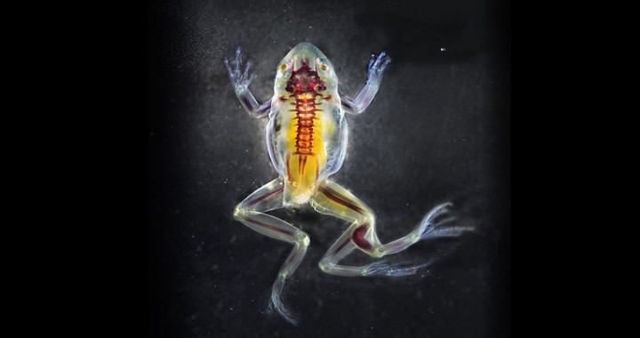
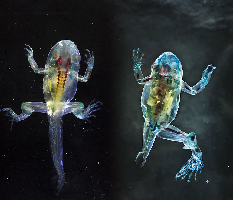
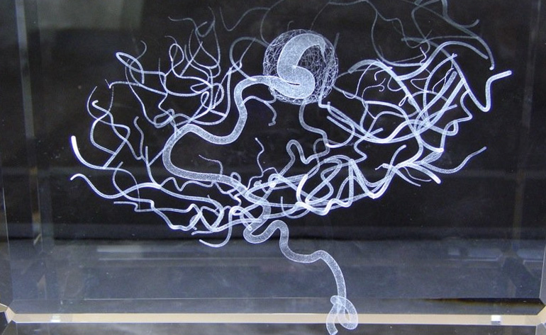
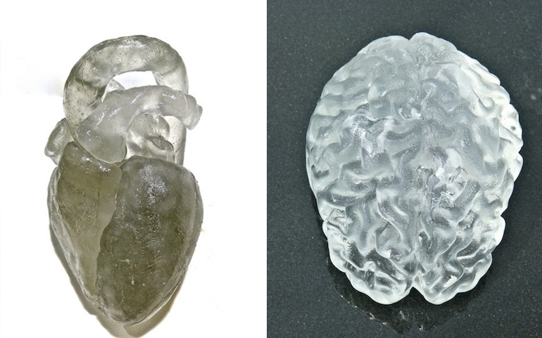
Greenberg combines art, science, and engineering to create microscopic photographs that reveal the hidden beauty of sand, flowers, food, and the human body. Greenberg builds the tools of his craft; he invented the “high-definition, three-dimensional light microscopes” with which he creates his photographs.1 His images give us unfamiliar and intimate views of materials we encounter every day.

Greenberg's Body Images
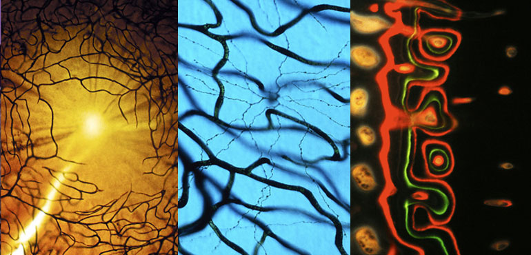
Microscopic images of retinas and bone.

Greenberg's Flower Images
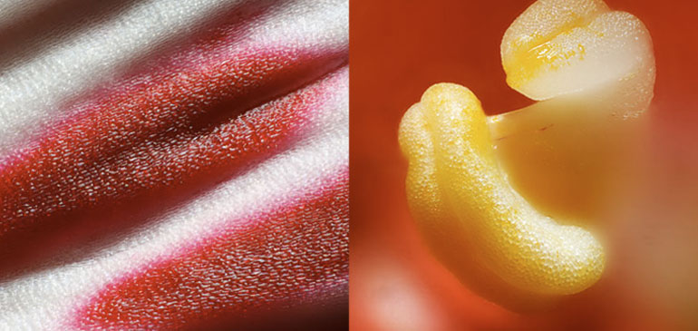
Microscopic images of Royal Poinciana and Hibiscus flowers.

See Greenberg’s site.

1See Greenberg’s biography.

BRANDON BALLENGEE

For twenty years, Ballengee has studied and documented deformed amphibians. He creates portraits of the deceased animals using a biology-lab process that removes everything but the collagen and stains the bones and cartilage. Ballangee sees his portraits as a way to memorialize the animals. He states: “If we start to look at the environment as made up of individuals just as unique as each and every one of us, I think that has the potential to really reframe our approach towards our own actions every day.”1

See Ballengee’s website.

Brandon Ballengee

1 Annie Minoff, “SciArts Spotlight: Brandon Ballengee,” (Science Friday: April 4, 2014), www.sciencefriday.com/blogs/04/04/2014/sciarts-spotlight-brandon-balleng-e.html?series=20.

TOBIE KERRIDGE, NIKKI STOTT, and IAN THOMPSON

Kerridge and Stott create speculative design projects that experiment with new materials.

Biojewellery, 2003-7
Working with bioengineer Ian Thompson, Kerridge and Scott created wedding bands coated with bone cells. “The bone tissue was cultured in a laboratory and then seeded onto a bioactive ceramic that acted as a scaffold for the growing cells….The final bone tissue was taken to the designer’s studio and combined with precious metals to finish the ring.”1 The ring functions as both a symbol of commitment and an actual memento of your partner’s corporeality.

Biojewellery

See the Biojewellery website.

1 Paola Antonelli, ed., Design and the Elastic Mind.(New York: The Museum of Modern Art, 2008), 111.

KATHARINE DOWSON

Dowson creates portraits of the brain and vascular system using laser-etched glass.

Memory of Brain Malformation, 2006
Dowson recreates a blood-flow x-ray of her cousin’s brain. The x-ray reveals a tumor that is later removed.1

Memory of Brain Malformation, Katharine Dowson

My Brain and My 3D Heart, 2013
Dowson references MRI scans of her brain and heart to create 3D printed versions of those organs. She then casts the organs in glass.

My Brain, My 3D Heart, Katharine Dowson

See Downson’s website.

1 Arthur I. Miller, Colliding Worlds: How Cutting-Edge Science is Redefining Contemporary Art (New York: W. W. Norton and Company, 2014), 192.

SUSAN ALDWORTH

Aldworth layers hand-drawn images onto the documents of medical science. Her work humanizes the dehumanized images of pathology; she reveals the subject in our maps of anatomy.

Cogito Ergo Sum 3, 2006
Aldworth combines fMRI scans of her brain with expressive imagery.

Cogito Ergo Sum 3, Susan Aldworth

See Aldworth’s website.

EDWARD BURTYNSKY

Burtynsky photographs landscapes that highlight the scale of human industry and consumption.

“My work does become a kind of lament....We can't have our cities, we can't have our cars, we can't have our jets without creating wastelands. For every act of creation there is an act of destruction. Take the skyscrapper—there is an equivalent void in nature: quarries, mines."1

See Burtynsky's website.

1Diane Ackerman, The Human Age: The World Shaped by Us (New York: W. W. Norton and Company, 2014), 24.